
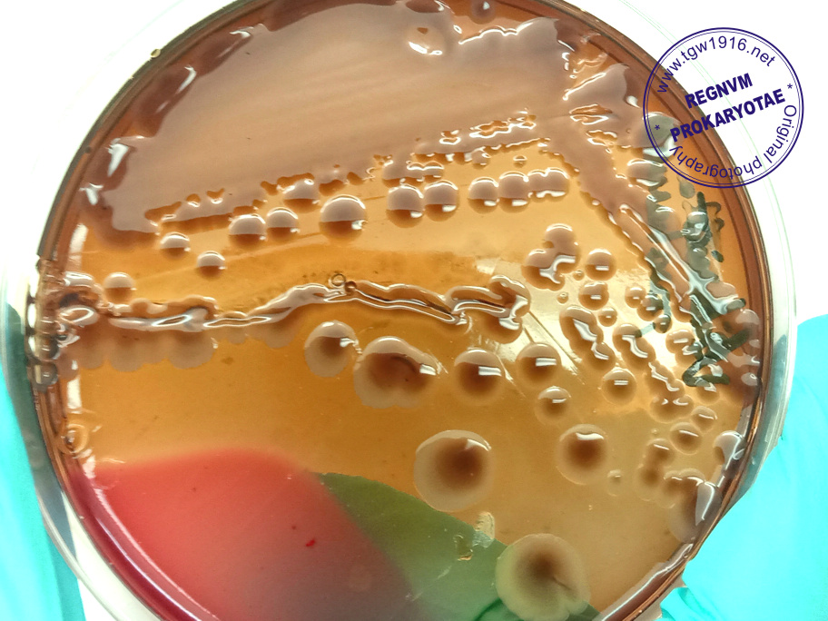
| Dark-center colonies and huge hemolysis area after 7 days incubation at room temperature |
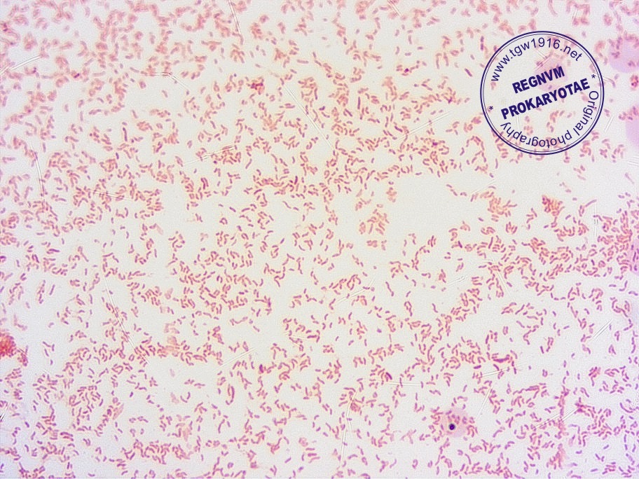
| V. metschnikovii Gram-negative, curved rods |
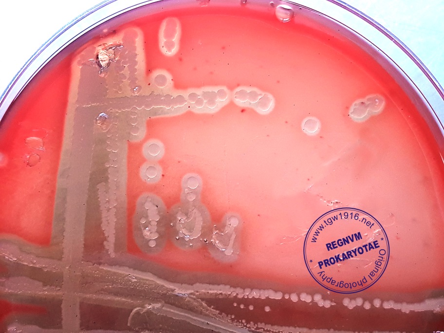
| Vibrio metschnikovii beta-hemolytic colonies |
| Vibrio metschnikovii |
Taxonomy
Morphology
Cultural characteristics
Biochemical characters
Ecology
Pathogenicity
References
Phylum Pseudomonadota (Proteobacteria), Class Gammaproteobacteria, Order Vibrionales, Family Vibrionaceae, Genus Vibrio,
Vibrio metschnikovii Gameleia 1888.
Historical synonym: Vibrio cholerae biovar proteus Shewan and Veron 1974.
Vibrio metschnikovii Gameleia 1888.
Historical synonym: Vibrio cholerae biovar proteus Shewan and Veron 1974.
Gram-negative, straight or curved rods, 0.6-0.8 x 2-3 µm, variable motility. No lateral
flagella on solid media.
flagella on solid media.
Beta-hemolytic on T-soy agar with 5% sheep blood. Opaque, glistening, dark-greyish,
flattened, convex colonies, pale-grey centers, intact edges, diameter 3-4 mm on blood
agar in 24 hours. Hemolysis enhances after prolonged incubation at 25 ºC (admin
note). Can grow in nutrient broth with: 1% NaCl & 6% NaCl. Variable growth in 8%
NaCl. No growth in 0% NaCl, 10% NaCl & 12% NaCl. Can grow at pH 10. Optimal
growth at 30-40 ºC. At 4 ºC grow slowly but become non-viable or non-culturable.
flattened, convex colonies, pale-grey centers, intact edges, diameter 3-4 mm on blood
agar in 24 hours. Hemolysis enhances after prolonged incubation at 25 ºC (admin
note). Can grow in nutrient broth with: 1% NaCl & 6% NaCl. Variable growth in 8%
NaCl. No growth in 0% NaCl, 10% NaCl & 12% NaCl. Can grow at pH 10. Optimal
growth at 30-40 ºC. At 4 ºC grow slowly but become non-viable or non-culturable.
Isolated from fresh, brackish and marine waters, sewage, cockles, shrimp, lobster,
crab and fowl, rarely from human clinical specimens (blood, urine, a foot wound, gall
bladder and other sources). One isolate from the lung of a dead calf.
A strain isolated from rooster bone marrow was resistant to O/129 vibriostatic agent
(admin note).
crab and fowl, rarely from human clinical specimens (blood, urine, a foot wound, gall
bladder and other sources). One isolate from the lung of a dead calf.
A strain isolated from rooster bone marrow was resistant to O/129 vibriostatic agent
(admin note).
First time has been isolated from a fowl that had died of a “cholera-like” disease.
Isolated from blood from an woman (82 year old) with peritonitis and an inflamed
gallbladder. May produce pneumonia, wound infections, bacteremia and septicaemia.
Isolated from blood from an woman (82 year old) with peritonitis and an inflamed
gallbladder. May produce pneumonia, wound infections, bacteremia and septicaemia.
- J.J. Farmer, M. Janda, 2004. Family I. Vibrionaceae. In: Bergey’s Manual of
Systematic Bacteriology, Second edition,Vol two, part B, George M. Garrity (Editor-
in-Chief), pp. 491-546. - J. G.Holt et al., 1994. Group 5 Facultatively anaerobic Gram-negative rods.
Subgroup 2 Family Vibrionaceae. In: Begey’s Manual of Determinative
Bacteriology, 9-th edition, Williams & Wilkins. pp. 190-194. - Judith A. Johnson, 2006. Vibrio. In: Topley & Wilson’s Microbiology and Microbial
Infections, 10 edition, Vol. 2, Bacteriology, Edward Arnold Ltd., 1507-1523. - Hansen, W, Freney, J, Benyagoub, H, Letouzey, M N, Gigi, J, Wauters, G. Severe
human infections caused by Vibrio metschnikovii.J. Clin. Microbiol. 1993 31: 2529-
2530.
Positive results for: methyl red (1% NaCl), Voges-Proskauer (1% NaCl), gelatin
hydrolysis (1% NaCl, 22 ºC), lipase, catalase, acid production from D-glucose,
glycerol, maltose, trehalose, D-mannitol, D-mannose and sucrose.
Negative results for indole production (Heart Infusion Broth, 1% NaCl), H2S on TSI,
urea hydrolysis, phenylalanine deaminase, ornithine (1% NaCl), nitrate reduction to
nitrite, oxidase, gas production from D-glucose, acid production from D-adonitol,
L-arabinose, D-arabitol, cellobiose, dulcitol, melibiose, raffinose, L-rhamnose, salicin
and D-xylose.
Variable results for citrate utilization (Simmons), arginine (1% NaCl), lysine (1%
NaCl), esculin hydrolysis, ONPG test, acid production from D-galactose, myo-inositol,
lactose and D-sorbitol.
hydrolysis (1% NaCl, 22 ºC), lipase, catalase, acid production from D-glucose,
glycerol, maltose, trehalose, D-mannitol, D-mannose and sucrose.
Negative results for indole production (Heart Infusion Broth, 1% NaCl), H2S on TSI,
urea hydrolysis, phenylalanine deaminase, ornithine (1% NaCl), nitrate reduction to
nitrite, oxidase, gas production from D-glucose, acid production from D-adonitol,
L-arabinose, D-arabitol, cellobiose, dulcitol, melibiose, raffinose, L-rhamnose, salicin
and D-xylose.
Variable results for citrate utilization (Simmons), arginine (1% NaCl), lysine (1%
NaCl), esculin hydrolysis, ONPG test, acid production from D-galactose, myo-inositol,
lactose and D-sorbitol.

(c) Costin Stoica


| Back |